Digital radiography uses electronic sensors and computer systems to capture detailed images of teeth, bone, and supporting structures. Unlike traditional film x-rays, digital sensors convert exposure into digital data instantly, allowing clinicians to view, enhance, and store images without waiting for chemical processing. This technological shift has reshaped routine diagnostics, making it easier for dental teams to assess conditions such as cavities, bone loss, and abnormalities with greater clarity.
For patients, the practical differences are immediately apparent: images appear on a monitor as soon as they are taken, and clinicians can adjust contrast, zoom in, and use measurement tools to evaluate the exact size and position of an issue. The speed and flexibility of digital files support more efficient appointments and clearer communication between the patient and care team. Images are also easier to archive and retrieve as part of a patient’s long-term record.
While the underlying physics of x-ray production remains consistent with conventional radiography, the digital workflow enhances diagnostic precision and clinical workflow. This convergence of sensor technology and software tools means dental teams can detect subtle changes earlier and plan restorative or preventive care with better information. In short, digital radiography is not just a different way of taking x-rays—it’s a platform that improves both the quality and usability of diagnostic imaging.
Contemporary digital sensors are engineered to capture a wide dynamic range of information, presenting fine gradations of density that film often cannot reproduce. These sensors come in two primary types—direct digital sensors and photostimulable phosphor plates—both of which offer superior detail resolution that benefits diagnosis of small fractures, early-stage decay, and periodontal bone changes. The result is clearer images that reduce ambiguity and support better clinical decisions.
Software tools that accompany these sensors add another layer of diagnostic power. Clinicians can apply filters, measure distances precisely, and compare images side-by-side to evaluate progression over time. Such capabilities are particularly valuable in prosthodontic planning and implant placement, where millimetric precision matters. The combination of high-resolution capture and robust image-processing tools elevates the diagnostic process beyond what was possible with film.
Because digital images can be standardized, they also improve consistency across appointments and practitioners. Standardized imaging protocols make it easier to repeat views with comparable results, which is crucial for monitoring healing, disease progression, or the performance of restorations. For patients, this translates into a more predictable and evidence-based approach to treatment planning.
One of the most tangible benefits of digital radiography is the immediacy of results. Images appear on-screen within seconds, allowing clinicians to evaluate findings in real time and discuss them with patients during the same visit. This instant feedback accelerates decision-making and reduces the need for follow-up appointments simply to review imaging. When time is valuable, a same-visit discussion supported by on-screen imagery improves understanding and fosters informed consent.
Digital imaging also enhances patient education. With tools to adjust contrast, annotate, and zoom in, clinicians can point out areas of concern in a way that is visually clear and easy to grasp. Patients who can see the problem and understand its context are better positioned to participate actively in their care. Modern dental practices use this capability to create collaborative treatment planning conversations rather than unilateral recommendations.
From a practice-efficiency standpoint, the digital workflow reduces administrative steps tied to film development, physical storage, and manual record-keeping. Images can be saved directly to an electronic health record, retrieved instantly for future visits, and shared with specialists or labs when collaborative care is needed, all without the delays and degradation risks associated with physical films.
Patients often ask about safety, and it’s important to address radiation exposure with clear, evidence-based information. Digital radiography typically requires a lower radiation dose than traditional film x-rays to achieve comparable or superior image quality. Technological advances in sensor sensitivity and image processing are central to this reduction, allowing clinicians to obtain diagnostic images while minimizing exposure.
Beyond the inherent efficiency of digital capture, contemporary practices follow established safety protocols to further reduce risk. Use of rectangular collimation, positioning aids, and lead aprons when appropriate ensures exposures are limited to the smallest practical area. Clinicians also adhere to professional guidelines that recommend imaging only when clinically justified and at appropriate intervals, balancing diagnostic benefit with patient safety.
For patients with specific concerns—such as pregnancy, previous high radiation exposure, or certain medical conditions—clinicians can explain why a particular image is necessary and what precautions will be taken. Transparent communication about radiation, paired with visible safety measures, helps patients feel informed and reassured about the imaging process.
Digital radiographs integrate seamlessly with modern practice management systems and digital treatment workflows. When images are part of an electronic health record, they can be accessed alongside clinical notes, intraoral photographs, and 3D scans, creating a unified clinical dataset that supports comprehensive care. This integration is especially important for complex restorative cases, implant planning, and long-term periodontal monitoring.
Because digital files are easily shared, collaboration with specialists, laboratories, or referring dentists becomes straightforward. Securely transmitting images—along with clinical notes—helps ensure that everyone involved in a patient’s care is working from the same detailed information. This coordinated approach reduces the potential for miscommunication and streamlines case planning across multiple providers.
Finally, the environmental and operational benefits of digital systems are notable. Eliminating chemical developers and film reduces hazardous waste and the associated handling risks, while digital archives require less physical space and are less prone to loss or deterioration. Together, these advantages make digital radiography a practical, sustainable choice for modern dental care.
In summary, digital radiography represents a significant advance in dental imaging: it improves image quality, speeds diagnosis, enhances patient communication, and supports safer, more collaborative care. The practice at The Haddon Dentist has adopted these technologies to provide patients with efficient, evidence-based diagnostics within a thoughtful and patient-centered workflow. If you would like to learn more about how digital imaging is used during your appointment or have questions about what to expect, please contact us for more information.


Digital radiography uses electronic sensors and computer systems to capture images of teeth, bone and supporting structures, converting x-ray exposure into digital data almost instantly. Unlike film-based x-rays, digital capture eliminates chemical processing and produces images on-screen within seconds for immediate review. This rapid conversion improves workflow and makes images easier to enhance, compare and archive.
Because images are digital files, clinicians can apply contrast adjustments, magnification and measurement tools that are not available with film, which improves visibility of subtle changes. The speed and flexibility of the digital workflow support more efficient appointments and clearer communication between patient and care team. Digital radiography therefore serves both diagnostic and practical needs in everyday dental care.
Contemporary digital sensors capture a wide dynamic range and higher detail resolution than traditional film, which helps identify small fractures, early decay and subtle bone changes. Sensors typically include direct digital detectors and photostimulable phosphor plates, both designed to deliver clearer images with less ambiguity. These hardware improvements reduce the chance of missed findings and support more confident clinical decisions.
Image-processing software further enhances diagnostic capability by enabling filters, precise measurements and side-by-side comparisons of serial images. Standardized imaging protocols combined with processing tools increase consistency across visits and practitioners, which is important for monitoring progression or healing. Together, sensor technology and software elevate the level of diagnostic information available for treatment planning.
Digital radiography typically requires a lower radiation dose than conventional film x-rays because modern sensors are more sensitive and software can optimize image quality from smaller exposures. Although the basic physics of x-ray generation remains the same, technological advances reduce the dose needed to obtain diagnostic images. This reduction, along with procedural safeguards, helps minimize patient exposure while preserving image quality.
Clinicians follow established safety measures such as rectangular collimation, positioning aids and the use of protective aprons when appropriate to limit exposure to the smallest practical area. Professional guidelines recommend imaging only when clinically justified and at appropriate intervals to balance diagnostic benefit and safety. When special circumstances arise, providers will explain the rationale for imaging and any additional precautions taken.
Digital radiographs play a central role in assessing tooth structure, root anatomy, periodontal bone levels and periapical health, all of which inform restorative decisions. High-resolution images help clinicians evaluate the extent of decay, the fit of existing restorations and the quality of supporting bone before restorative procedures. For implant planning, millimetric detail in two-dimensional images complements three-dimensional scans to define implant position and angulation with greater confidence.
When combined with digital impressions and CAD/CAM workflows, radiographic data becomes part of an integrated treatment plan that guides surgical and prosthetic steps. Clear imaging reduces uncertainty during restoration and implant placement, supporting predictable, evidence-based outcomes. This coordinated approach improves both function and long-term prognosis of restorations.
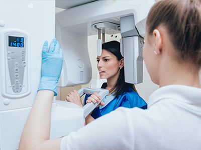
During a digital radiography appointment a sensor or plate is positioned in or around the mouth and an exposure is made; the process takes only a few seconds and causes little to no discomfort. The resulting images appear on a monitor immediately, allowing the clinician to confirm image quality on the spot and retake views if necessary. Positioning aids and bite blocks are used to achieve consistent, diagnostic images with minimal repeats.
After acquisition the clinician will often review images with you using on-screen tools to point out areas of interest, compare with prior studies and explain recommended next steps. This instant review supports same-visit discussions and helps you participate in treatment decisions. If additional views or 3D imaging are needed, the care team will explain the reasons before proceeding.
Digital radiographs are saved directly to an electronic record and archived in a standardized format, which makes retrieval fast and reliable for future visits. Electronic storage reduces the physical space and degradation risks associated with film and simplifies organization of long-term records. Images can be indexed and linked to clinical notes, intraoral photos and other diagnostic data for a comprehensive patient chart.
Patient images and records are managed within secure, HIPAA-informed systems that use access controls, regular backups and audit trails to protect privacy and data integrity. The Haddon Dentist employs industry-standard safeguards to limit access to authorized staff and to maintain secure transmissions when images are shared. These protections help ensure that diagnostic information is preserved and accessed only as clinically appropriate.
Yes, one of the practical advantages of digital files is that they can be securely transmitted to specialists, laboratories or referring providers without the delays of physical film. Shared images can include annotations and comparative views, which improves clarity when discussing complex cases. Electronic sharing supports coordinated care by ensuring all clinicians work from the same diagnostic dataset.
Secure transfer reduces duplication of imaging and speeds collaborative planning, particularly for implant cases, surgical consultations and complex restorative work. When sharing is required, clinicians include relevant clinical notes to give receiving providers proper context. This streamlined exchange supports more efficient and consistent patient care across providers.
Digital radiography is commonly used for pediatric patients with size-appropriate sensors and protocols that reduce exposure while maintaining diagnostic quality. Clinicians tailor imaging techniques to the child’s size and clinical need, using shielding and positioning aids to minimize repeats and limit dose. Regular preventive imaging schedules are adjusted to balance early detection with radiation stewardship for growing patients.
For pregnant patients, dental imaging is performed only when clinically necessary and with additional precautions such as lead aprons and thyroid collars to limit exposure. Providers discuss the risks and benefits and defer nonurgent imaging when appropriate, coordinating care with the patient’s medical team if needed. Transparent communication and conservative protocols help ensure safety while addressing urgent oral health concerns.
Digital images on-screen give clinicians tools to highlight anatomy, annotate findings and show side-by-side comparisons that make complex conditions easier to understand. When patients can see a clear image and a clinician’s annotated explanation, they are better positioned to comprehend treatment needs and alternatives. Visual evidence supports informed consent and reduces uncertainty about recommended care.
These capabilities foster collaborative treatment planning, where patients participate in choosing options based on clearer information and realistic expectations. Serial imaging also enables tracking of disease progression or healing, so patients can see objective results over time. This transparency strengthens the clinician–patient relationship and supports long-term oral health goals.
Digital radiography is embedded in modern treatment workflows by linking images to electronic health records, digital impressions and CAD/CAM systems to create a unified clinical dataset. This integration streamlines same-visit diagnostics and restorative procedures, reduces administrative steps tied to film processing and supports real-time collaboration among the care team. Standardized imaging protocols help maintain consistency across appointments and providers.
The Haddon Dentist has adopted these digital tools to support evidence-based diagnostics and efficient restorative workflows, enabling clinicians to plan and explain care with precision. Combining radiographs with intraoral photography and digital impressions helps the practice deliver predictable outcomes while keeping patients informed throughout their treatment journey.

The path to a restored, high-functioning smile is a journey of precision, and it begins with a single, focused conversation. We invite you to experience the intersection of clinical mastery and uncompromising luxury at The Haddon Dentist. Whether you are seeking the immediate results of CEREC® same-day crowns, exploring the life-changing benefits of dental implants, or ready to revitalize your aesthetic with professional teeth whitening, we are here to curate a plan tailored exclusively to you. Your time is your most valuable asset, and we cherish the opportunity to provide the elite care you deserve in an environment that respects your comfort and your goals.