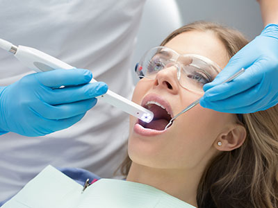
An intraoral camera is a compact, pen-sized imaging device designed specifically for use inside the mouth. Unlike traditional external photography, this tool captures high-resolution, full-color images of teeth, gums, and other oral tissues from angles that would otherwise be difficult to visualize. Its small tip and maneuverable head allow clinicians to obtain detailed close-ups of surfaces, margins, and soft-tissue conditions while the patient remains comfortable in the chair.
Technically, the device combines miniature optics, a bright LED light source, and a digital sensor to produce instant stills or live video that can be displayed on a monitor. Modern intraoral cameras deliver crisp images with accurate color rendering and sharp detail, making it possible to see hairline fractures, early enamel defects, and subtle tissue changes that might be missed on a cursory exam.
For patients, the intraoral camera transforms an abstract clinical observation into a clear visual. What once required descriptive language or indirect explanation can now be shown directly, which makes examinations more transparent and easier to understand. This basic shift—from telling to showing—is at the heart of why intraoral imaging has become a standard component of modern dental care.
When clinicians use an intraoral camera, the conversation with the patient becomes collaborative rather than one-sided. Instead of relying solely on verbal descriptions or X-ray interpretation, the provider can place the camera, display the image on a screen, and walk the patient through what is visible. This shared viewing experience helps patients comprehend the nature and extent of oral conditions, which supports informed decision-making and reduces uncertainty.
Visual evidence also supports educational moments during routine checkups. Dentists can point out the early stages of wear, the locations of plaque accumulation, or the precise margins of a restorative material, and then demonstrate proper brushing and flossing techniques in context. Seeing the problem makes preventive advice more tangible and actionable for patients of all ages.
Moreover, intraoral imaging fosters trust by documenting the baseline condition and tracking progress over time. Patients appreciate seeing their own anatomy up close; it removes guesswork and empowers them to participate in treatment planning. For clinicians, this clearer dialogue can lead to more productive conversations and better long-term adherence to recommended care plans.
From a diagnostic standpoint, the intraoral camera enhances the clinician’s ability to detect problems early. Enamel craze lines, micro-fractures, marginal breakdown around restorations, and subtle mucosal lesions are far easier to identify when magnified and illuminated by a camera. Early detection often means more conservative options can be considered, preserving tooth structure and simplifying future care.
Documentation is another major benefit. Individual images captured during an appointment become part of the patient’s chart and serve as objective records that can be referenced later. These images are invaluable when monitoring suspicious areas, comparing pre- and post-treatment outcomes, or communicating findings to specialists or laboratories. Clear photographic records improve continuity of care and help ensure that everyone involved in a case is working from the same visual information.
When planning restorations or complex procedures, intraoral images allow the clinical team to review morphology and margins with precision. High-quality photos help the dentist evaluate occlusal relationships, identify preparation needs, and confirm the integrity of existing work. In combination with other diagnostic tools, intraoral cameras contribute to more predictable, well-documented treatment pathways.
Intraoral cameras are most powerful when they plug into a broader digital ecosystem. Contemporary dental practices integrate camera output directly into electronic health records, image management systems, and CAD/CAM workflows. This seamless connectivity streamlines case presentation, record keeping, and communication with dental labs or referral partners, eliminating the friction of manual image transfer and simplifying collaborative treatment planning.
Software enhancements allow clinicians to annotate images, compare side-by-side views from different visits, and adjust contrast or magnification to emphasize specific details. These digital tools make it easier to create visual narratives for patients and colleagues alike, and to store images in formats that are secure, searchable, and accessible during subsequent appointments.
Because intraoral cameras are compatible with many contemporary imaging platforms, they naturally complement digital impression systems like CEREC as well as conventional radiography. The combination of real-time clinical photography and three-dimensional scans gives clinicians a richer dataset for restorative and prosthetic work, increasing the precision of laboratory communication and in-office fabrication alike.
Having images taken with an intraoral camera is quick and noninvasive. During the exam, the clinician gently guides the device over the teeth and soft tissues while the patient remains seated comfortably. Most people experience no discomfort beyond the normal minor sensations associated with an oral exam; the process typically adds only a few minutes to a standard visit but provides significant diagnostic value.
Infection control and patient safety are central to clinical use. Tips and sleeves designed for intraoral cameras are disinfected or single-use, and clinicians follow established sterilization protocols to prevent cross-contamination. The hardware itself is robust and designed for frequent clinical use, while software safeguards protect patient privacy by limiting access to stored images within the practice’s secure records system.
After images are captured, the clinician will usually review them with the patient and explain the findings, recommended next steps, and any monitoring plan. Because images are stored in the chart, they can be revisited at follow-up visits to document healing, verify the performance of restorations, or assess the effectiveness of preventive measures. This continuity makes the intraoral camera an ongoing asset to both patient education and clinical oversight.
Overall, the integration of intraoral imaging into routine care elevates diagnostic clarity and patient engagement without adding complexity to the appointment. When applied thoughtfully, it becomes a practical tool that supports conservative treatment, better communication, and measurable clinical outcomes.
At The Haddon Dentist, we incorporate intraoral imaging into comprehensive care to help patients see—and understand—their oral health more clearly. If you’d like to learn how intraoral camera technology is used during examinations or how it might affect your treatment planning, please contact us for more information.


An intraoral camera is a compact, pen-sized imaging device designed to capture high-resolution, full-color photos and live video of teeth, gums and other oral tissues. It combines miniature optics, a bright LED light source and a digital sensor to produce detailed close-ups from angles that are difficult to see with the naked eye. Clinicians can display the images on a monitor instantly, turning small findings into clear visuals for diagnosis and education.
Modern intraoral cameras deliver accurate color rendering and high magnification, which make it easier to identify hairline fractures, early enamel defects and subtle soft-tissue changes. Because the device records stills and video, images can be stored in the patient record for comparison over time. This capability has made intraoral imaging a routine component of contemporary dental examinations.
When clinicians use an intraoral camera, the exam becomes a shared visual experience rather than a one-sided description. The dentist can display live images on a screen and walk the patient through what is visible, which improves understanding and supports informed decision making. Visual evidence also makes preventive instruction more practical by showing plaque, wear patterns and the margins of restorations in context.
At The Haddon Dentist we incorporate intraoral imaging into consultations to help patients see the exact issue and discuss conservative treatment options when appropriate. This transparency supports trust and helps patients participate actively in their care plan. Images captured during the visit can also be annotated to clarify findings for future reference.
Intraoral cameras enhance the detection of early-stage problems that may be missed on a cursory exam, including enamel craze lines, micro-fractures, marginal breakdown around restorations and subtle mucosal lesions. The magnified view and high illumination improve visualization of surface texture, margins and color changes that warrant monitoring or intervention. Detecting these issues early often allows for more conservative treatment and better long-term outcomes.
Cameras are also helpful for documenting progression of wear, the effectiveness of hygiene routines and the fit of existing restorations. When combined with other diagnostics like radiographs or digital impressions, intraoral images contribute to a fuller clinical picture. Clinicians can use this combined dataset to prioritize care based on objective visual evidence.
Yes; intraoral imaging is quick, noninvasive and typically well tolerated by patients of all ages. The clinician gently guides the pen-sized device over teeth and soft tissues while the patient remains seated, and the process usually adds only a few minutes to a routine visit. Single-use sleeves or disinfected tips help minimize any contact concerns.
Infection control protocols are followed for every use, including barrier sleeves and sterilization procedures for reusable components when required. Software safeguards keep images secure within the electronic health record and limit access to authorized team members. Together, these measures make intraoral imaging a safe and practical addition to dental care.
High-quality photographs become part of the patient chart and serve as objective records for diagnosis, treatment planning and follow-up. Images allow the clinical team to evaluate tooth morphology, margins and occlusal relationships more accurately when planning restorations or complex procedures. Clear photographic documentation also improves communication with dental laboratories and referral partners.
Stored images can be compared visit to visit to monitor healing, verify restoration performance and document the need for future intervention. Annotation tools and side-by-side comparisons make it possible to create a visual narrative that supports clinical decisions. For clinicians, this documentation reduces ambiguity and helps maintain continuity of care.
Intraoral cameras are designed to plug into modern digital ecosystems, allowing images to be stored in electronic health records, integrated with image management systems and linked to CAD/CAM workflows like CEREC. This interoperability streamlines case presentation and laboratory communication by keeping visual data centralized and accessible during treatment. Software features often let clinicians annotate images and compare them directly with digital impressions.
In practices that use same-day crown systems, such as those offering CEREC restorations, intraoral images complement three-dimensional scans by providing real-time clinical photographs of margins and soft tissues. At The Haddon Dentist this combined dataset contributes to efficient, well-documented restorative workflows that prioritize precision and patient understanding. The result is a more predictable process when fabricating both in-office and laboratory restorations.
Images captured with an intraoral camera are uploaded to the practice’s secure records system and become part of the permanent chart. Electronic health records protect access through user permissions and encryption, helping ensure that only authorized providers and staff can view sensitive images. Secure storage also makes the images searchable and easy to retrieve during subsequent visits.
Clinicians may annotate or compare images from different dates to monitor changes or document treatment outcomes. When images are shared with specialists or laboratories, they are transmitted through secure, HIPAA-compliant channels whenever required. These safeguards balance accessibility for clinical care with the privacy protections patients expect.
Yes; clinicians routinely review intraoral images with patients during the appointment so findings are clear and questions can be answered immediately. Seeing their own anatomy up close helps patients understand the nature and extent of conditions and the rationale behind recommended care. This review is an educational opportunity to demonstrate targeted home care techniques where appropriate.
After the review, images remain in the chart for future reference and can be revisited at follow-up visits to document healing or restoration performance. Clinicians can annotate specific areas on the images to highlight points of concern or progress. Patients are encouraged to ask questions so they leave the appointment with a clear understanding of next steps.
The frequency of intraoral imaging depends on individual risk factors, the presence of existing restorations and clinical findings during the exam. For many patients, capturing images at annual checkups or when a specific concern is identified provides useful baseline documentation and a reference for monitoring changes. Patients with high caries risk, extensive restorations or prior oral pathology may benefit from more frequent imaging.
Clinicians use their professional judgment to determine when images add diagnostic or educational value, balancing clinical need with appointment time. Because the process is fast and noninvasive, it can be incorporated selectively without disrupting routine care. Discussing your personal history and concerns with the dentist will help determine an appropriate schedule for imaging.
Trained members of the clinical team, including dentists, hygienists or certified assistants, typically perform intraoral imaging as part of the exam. These team members follow standardized techniques to capture consistent, diagnostic-quality views that support accurate assessment. If any particular views are requested or special documentation is needed, the clinician will communicate those requirements before imaging.
Capturing intraoral images usually adds only a few minutes to a standard appointment, since the device is quick to position and captures instant stills or video. The time investment yields valuable diagnostic information and visual records that streamline subsequent discussions and treatment planning. Patients should expect a brief, comfortable experience that enhances the clarity of their exam.
The path to a restored, high-functioning smile is a journey of precision, and it begins with a single, focused conversation. We invite you to experience the intersection of clinical mastery and uncompromising luxury at The Haddon Dentist. Whether you are seeking the immediate results of CEREC® same-day crowns, exploring the life-changing benefits of dental implants, or ready to revitalize your aesthetic with professional teeth whitening, we are here to curate a plan tailored exclusively to you. Your time is your most valuable asset, and we cherish the opportunity to provide the elite care you deserve in an environment that respects your comfort and your goals.