Oral and oropharyngeal cancers remain a significant public health concern because they can be life‑altering if not identified early. While these cancers make up a modest proportion of all malignancies, the consequences of delayed diagnosis are serious: more extensive treatment, greater impact on speech and swallowing, and reduced survival. Recent decades have seen shifts in the pattern of disease, with some types declining while HPV‑associated cases have risen, especially among younger adults.
Certain factors raise an individual’s chance of developing oral cancer. Tobacco use and frequent heavy alcohol consumption remain two of the most consistent contributors. Men historically face higher rates than women, and risk increases with age, particularly after 50. Occupational exposures, a poor diet, and a history of head and neck radiation also contribute to elevated risk for some patients.
Human papillomavirus (HPV), specifically high‑risk strains that affect the oropharynx, has changed the landscape of oral cancer in recent years. HPV‑linked cancers often occur in the tonsils and the base of the tongue and can affect people who do not have traditional risk factors such as smoking. Understanding these trends helps clinicians tailor screening and education to the needs of each patient.
Oral cancer can develop anywhere inside the mouth and in adjacent structures of the throat. Common sites include the mobile tongue, the floor of the mouth beneath the tongue, the gums, the inner lining of the cheeks, the hard palate, and the oropharynx (including the tonsils and base of tongue). Lesions in different locations can present with subtly different symptoms, which is why a systematic visual and tactile exam is essential.
Early signs are often painless and may be easy to overlook. Persistent white or red patches, unexplained sores that do not heal within two weeks, thickened or lumped tissue, or an area that looks different from surrounding mucosa should prompt further evaluation. Other clues can include persistent hoarseness, a change in voice, difficulty swallowing, unexplained ear pain, or a new lump in the neck.
Because early lesions can be subtle, patients should report any oral changes that persist beyond a short period. Routine oral cancer screening at dental visits is designed to catch abnormalities before they grow or spread, which offers the best chance for simpler, more effective treatment and a better prognosis.
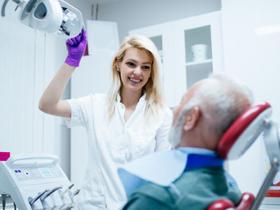
An oral cancer screening is a focused, noninvasive part of a comprehensive dental exam. The clinician begins with a review of your medical and dental history to identify risk factors like tobacco or alcohol use, prior radiation therapy, or symptoms you may have noticed. This background helps guide the exam and determine whether any additional tests are warranted.
The hands‑on portion of the screening consists of a careful visual inspection of the lips, gums, tongue, inner cheeks, floor and roof of the mouth, and the back of the throat when accessible. The clinician will also palpate the tissues inside the mouth and feel along the lower jaw and neck for any lumps or enlarged lymph nodes. This combination of sight and touch helps detect irregularities that may not be visible alone.
Some practices use adjunctive aids to support the clinical exam, such as special lights or screening tools that can highlight suspicious tissue. These technologies do not replace a thorough clinical assessment but can help identify areas that deserve closer attention. If an abnormality is found, your dentist will explain the findings and recommend a clear next step, which may include monitoring, referral for biopsy, or collaboration with a specialist.
Finding a suspicious lesion does not automatically mean cancer; many oral conditions are benign or related to irritation, infection, or autoimmune disease. However, any persistent lesion that cannot be explained by an identifiable cause requires further evaluation. The usual next step is referral to an oral surgeon, ENT specialist, or other head and neck specialist for definitive assessment.
Definitive diagnosis typically involves a biopsy, during which a small sample of tissue is taken and examined microscopically. Imaging studies may also be used to assess the extent of disease if cancer is suspected. These diagnostic steps help determine the stage and nature of the lesion so that an appropriate, evidence‑based treatment plan can be developed in collaboration with a multidisciplinary team.
Timely communication between dental and medical providers is important after an abnormal finding. A coordinated approach ensures that patients receive accurate diagnosis and prompt access to specialists when needed. Early referral and intervention can greatly expand treatment options and improve outcomes.
Prevention and early detection go hand in hand. Reducing modifiable risks—quitting tobacco, limiting alcohol intake, protecting lips from excessive sun exposure, and maintaining a nutritious diet—can lower the chance of developing oral cancer. Vaccination against HPV is an effective preventive measure for HPV‑related oropharyngeal disease and is an important public health tool for eligible individuals.
The dental team plays a central role in ongoing oral health surveillance. Regular dental visits offer repeated opportunities to detect change over time, reinforce preventive habits, and counsel patients about risk reduction. For those with prior oral cancer or with higher risk, more frequent examinations and a personalized surveillance plan are appropriate.
At The Haddon Dentist, our approach emphasizes thorough screening and clear communication. We work to ensure patients understand what was observed during an exam, why certain follow‑up steps are recommended, and how to recognize warning signs between visits. Collaboration with medical specialists is a routine part of our process when further assessment or treatment is indicated.
Oral cancer screening is a straightforward but powerful tool for protecting oral and overall health. By understanding risk factors, recognizing early signs, and incorporating routine examinations into regular dental care, patients and clinicians together improve the likelihood of early detection and better outcomes.
If you have questions about oral cancer screening or are concerned about changes in your mouth, please contact us for more information. Our team can explain the screening process, discuss risk factors, and guide you toward the appropriate next steps.


An oral cancer screening is a focused, noninvasive assessment performed as part of a routine dental exam to look for early signs of cancer in the mouth and oropharynx. The exam combines a visual inspection of the lips, gums, tongue, cheeks, floor and roof of the mouth and a tactile evaluation of the tissues and neck to detect lumps or abnormalities. Because early lesions are often painless, screening aims to identify subtle color changes, patches or masses before symptoms progress.
When indicated, the clinician may supplement the exam with targeted questions about symptoms, risk factors and recent changes in oral health. Screening can also include the use of adjunctive aids that help highlight suspicious tissue, though these tools do not replace a careful clinical evaluation. The overall goal is early detection so that any necessary follow-up can be started promptly and coordinated with appropriate specialists.
Several factors raise a patient’s risk for oral and oropharyngeal cancers, including long-term tobacco use in any form and frequent heavy alcohol consumption. Age is also a factor, with risk rising after about age 50, and men historically show higher incidence rates than women. Occupational exposures, a history of head and neck radiation, poor nutrition and certain chronic irritations can further increase risk for some patients.
Human papillomavirus, particularly high-risk strains that affect the oropharynx, has changed the risk profile by increasing cases among younger adults and people without traditional risk factors. Because risk varies by individual, a thorough medical and dental history helps the dental team tailor screening frequency and counseling. Patients should share information about tobacco, alcohol, sexual history relevant to HPV exposure and prior radiation when asked so the clinician can assess risk accurately.
The screening begins with a review of the patient’s medical and dental history to identify risk factors and symptoms that may need focused attention. The hands-on portion involves a systematic visual inspection of all oral tissues followed by palpation of the intraoral tissues and felt evaluation of the jaw and neck for enlarged lymph nodes or masses. This combination of sight and touch helps identify lesions that might be missed by visual inspection alone.
If the clinician finds an area of concern, they will document its size, location and appearance and discuss next steps with the patient. Depending on findings, recommended steps may include short-term monitoring, adjunctive screening aids, referral for biopsy or consultation with an oral surgeon or ENT specialist. Clear communication about observations and the rationale for referral is an important part of the visit.
Patients should notify their dental team about any oral changes that persist longer than two weeks, including unexplained white or red patches, sores that do not heal, thickened or indurated tissue and any new lumps. Other symptoms that warrant attention include persistent hoarseness, a change in voice, difficulty swallowing, ear pain without an ear problem and an unexplained neck mass. Early lesions are often painless, so attention to subtle changes is important.
Keeping a simple record or taking a photograph of a suspicious area can help the clinician assess change over time and determine whether further investigation is needed. Prompt reporting of symptoms allows for timely evaluation and, if necessary, expedited referral for definitive diagnosis. Regular dental visits remain a cornerstone of surveillance because clinicians can detect changes that patients may not notice.
High-risk strains of human papillomavirus (HPV), most notably HPV-16, are linked to a rising number of oropharyngeal cancers, particularly those originating in the tonsils and base of tongue. HPV-related cancers often occur in patients without classic risk factors such as tobacco or heavy alcohol use, which has shifted screening and education priorities in recent years. Understanding HPV’s role helps clinicians communicate risk and tailor screening for individual patients.
Prevention includes HPV vaccination for eligible individuals, which reduces the risk of HPV-related disease, and counseling patients about behaviors that lower exposure risk. The dental team can reinforce public health guidance about vaccination and safe practices and incorporate HPV risk assessment into routine history-taking. When HPV-related cancer is suspected, coordinated care with medical specialists is essential for accurate diagnosis and treatment planning.
Finding a suspicious lesion does not mean a diagnosis of cancer, as many oral abnormalities are benign or related to infection, irritation or inflammatory conditions. The clinician will evaluate the lesion’s characteristics and may recommend observation for a short interval if the appearance suggests a benign cause, or they will refer the patient for further assessment if the lesion is persistent or shows concerning features. Referral is commonly made to an oral surgeon, otolaryngologist or other head and neck specialist for definitive evaluation.
Definitive diagnosis often requires a biopsy in which a small tissue sample is examined microscopically, and imaging may be used to assess extent if malignancy is suspected. Timely coordination between the dental practice and medical specialists helps ensure the patient receives an evidence-based diagnostic workup and prompt access to appropriate care. Clear explanations of the rationale for referral and next steps help patients understand the process and reduce uncertainty.
Adjunctive screening aids such as special lights, autofluorescence devices and toluidine blue can highlight tissue differences that are not obvious on routine inspection and may help clinicians identify areas that deserve closer scrutiny. These technologies are useful as supplemental tools but have limitations and cannot replace a careful clinical examination and clinical judgment. Sensitivity and specificity vary by device, and false positives or negatives can occur.
Because of these limitations, adjunctive tools are best used to guide further evaluation rather than as standalone diagnostic tests. When an adjunctive aid highlights an area of concern, the clinician will integrate that information with the clinical exam and history to decide whether to monitor, biopsy or refer. Patients should understand that adjunctive testing is one part of a broader diagnostic approach aimed at early and accurate detection.
Oral cancer screening is typically performed at routine dental checkups, which for many patients occur every six months as part of a comprehensive exam. Frequency may be increased for patients with higher risk factors, such as a history of tobacco or heavy alcohol use, prior head and neck radiation, a history of oral cancer or confirmed HPV exposure. The dental team will recommend a personalized schedule based on individual risk and clinical findings.
Patients at elevated risk may benefit from more frequent surveillance and a documented plan to track any suspicious changes over time. Routine visits provide repeated opportunities for early detection, reinforcement of preventive measures and timely referral when needed. Discuss your personal risk factors with your clinician so you and the team can agree on an appropriate screening interval.
The dental team is instrumental in primary prevention, early detection and long-term follow-up for patients at risk for oral cancer. Clinicians provide counseling on modifiable risks such as tobacco cessation, alcohol moderation, sun protection for the lips and the importance of a nutritious diet, and they reinforce public health measures like HPV vaccination for eligible patients. Regular dental visits allow the team to track tissue changes over time and to intervene early when abnormalities appear.
For patients with a history of oral cancer or those at higher risk, the practice can implement a personalized surveillance plan that includes more frequent exams, documentation of lesion appearance and coordination with medical specialists. Clear communication, timely referrals and collaborative care are central to improving outcomes, and the dental team acts as an ongoing partner in the patient’s oral and overall health. The Haddon Dentist emphasizes thorough screening and patient education as part of routine care.
The office of The Haddon Dentist combines specialist-level expertise and a systematic approach to screening within a modern clinical setting, offering patients thorough evaluations and clear explanations of findings. Our practice emphasizes an evidence-based process that integrates careful history-taking, meticulous clinical examination and, when appropriate, adjunctive tools or timely referral to head and neck specialists. Patients benefit from coordinated care and a focus on early detection to preserve function and optimize outcomes.
Choosing a dental team that prioritizes surveillance and patient education helps ensure changes are identified promptly and managed efficiently. We work to make the screening process understandable and to involve patients in decisions about follow-up and preventive strategies. If you have concerns or notice persistent changes in your mouth, contact the office so we can review your history and arrange an appropriate evaluation.

The path to a restored, high-functioning smile is a journey of precision, and it begins with a single, focused conversation. We invite you to experience the intersection of clinical mastery and uncompromising luxury at The Haddon Dentist. Whether you are seeking the immediate results of CEREC® same-day crowns, exploring the life-changing benefits of dental implants, or ready to revitalize your aesthetic with professional teeth whitening, we are here to curate a plan tailored exclusively to you. Your time is your most valuable asset, and we cherish the opportunity to provide the elite care you deserve in an environment that respects your comfort and your goals.